RelaxometryTools
Demonstrations.nb ››
Guide page ›› Code on Github ››
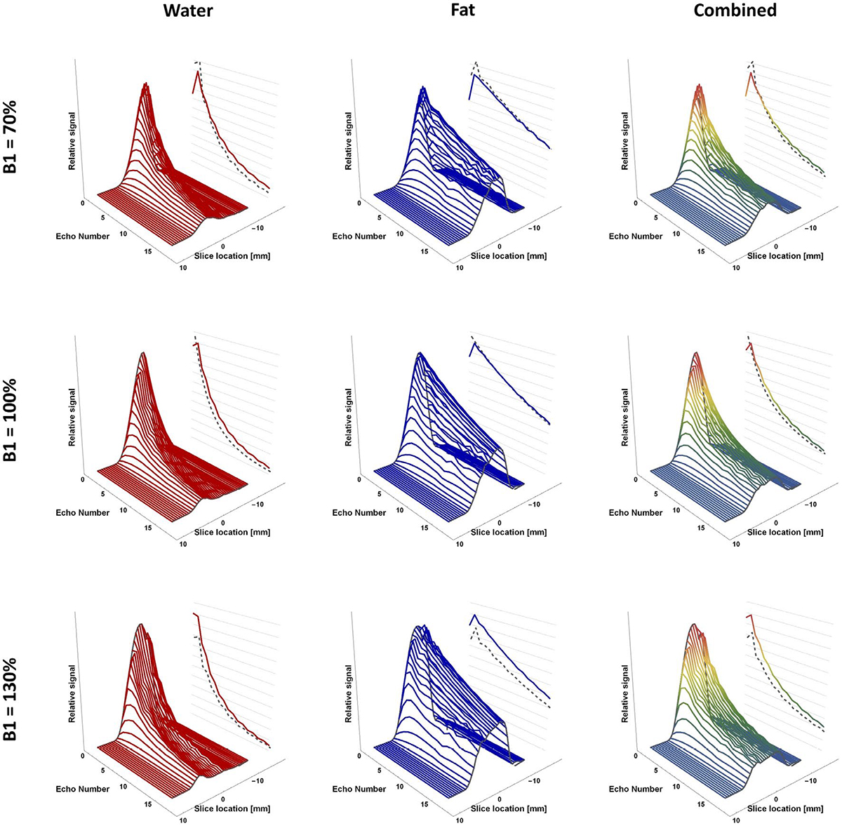
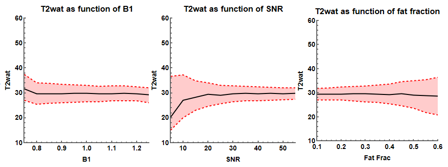
A collection of tools to fit T2, T2*, T1rho and T1 relaxometry data. The main function of this toolbox is an extended phase graph (EPG) (Weigel 2015) method for multi-compartment T2 fitting of multi-echo spin echo data (Marty et al. 2016). Therefore it provides functions to simulate and evaluate EPG (Keene et al. 2020). Back››


References
- Weigel, Matthias. 2015. “Extended phase graphs: Dephasing, RF pulses, and echoes - pure and simple.” Journal of Magnetic Resonance Imaging 41 (2). Wiley-Blackwell: 266–95. link››.
- Marty, Benjamin, Pierre Yves Baudin, Harmen Reyngoudt, Noura Azzabou, Ericky C. A. Araujo, Pierre G. Carlier, and Paulo L. de Sousa. 2016. “Simultaneous muscle water T2and fat fraction mapping using transverse relaxometry with stimulated echo compensation.” NMR in Biomedicine 29 (4): 431–43. link››.
- Keene, K. R., Beenakker, J. W. M., Hooijmans, M. T., Naarding, K. J., Niks, E. H., Otto, L. A. M., van der Pol, W. L., Tannemaat, M. R., Kan, H. E., and Froeling, M. “T2 relaxation-time mapping in healthy and diseased skeletal muscle using extended phase graph algorithms.” Magnetic Resonance in Medicine, 84(5), 2656–2670. link››